Aunque poco se habla de ello, el microambiente celular es determinante para la función celular normal y sus alteraciones podrían estar relacionadas con el desarrollo y empeoramiento de enfermedades (1,2). Cada vez es mayor el cuerpo de evidencia científica existente en torno a las alteraciones del microambiente celular y la perpetuación de la respuesta inflamatoria, muerte celular, desarrollo y progresión del cáncer y degeneración tisular (3–7). Así mismo, del microambiente celular adecuado depende también que puedan o no alcanzarse los mejores efectos terapéuticos en muchas enfermedades (8–11).
Microambiente celular: de lo normal a lo patológico
Cada una de las células del cuerpo se encuentra ubicada en una región particular y esta en contacto en ocasiones con más células y en otras con componentes de la matriz extracelular, hormonas, citoquinas, factores de crecimiento, otras moléculas de señalización intercelular, proteinas, vitaminas, minerales y otros nutrientes. A este conjunto se le denomina microambiente celular y no es único, existen múltiples como tejidos en el cuerpo (12–14).
Como resultado de la interacción y relación constante de las células y los diversos componentes de su microambiente se modifican su metabolismo, supervivencia, comunicación y función. En condiciones normales la función de los tejidos es resultado del fino ajuste en el microambiente celular (15–17).
Son varios los factores que modifican el microambiente celular, a continuación se mencionan algunos de ellos:
- Radiación ionizante (18)
- Ejercicio (19)
- Dieta (20)
- Inflamación crónica (21)
- Micronutrientes (22)
- Flujo sanguíneo (23)
- Hormonas (24)
- Estrés oxidativo (25)
- Microbiota intestinal (26)
La respuesta anormal de la célula ante un ambiente alterado
Indiscutiblemente la alteración del microambiente celular conduce al desarrollo de la enfermedad. El papel que tiene la alteración de este ambiente en el desarrollo de la enfermedad es un campo activo de investigación teniendo en cuenta las implicaciones que puede tener en términos terapéuticos. La medicina ortomolecular y la terapia con corticoides son ejemplos de terapéuticas dirigidas a modificar y normalizar el microambiente celular que se emplean con frecuencia en todo el mundo (27,28).
La exposición constante a factores que modifican el microambiente celular conduce a la alteración de la función celular y esto, a su vez, altera aún mas el microambiente. Un buen ejemplo es lo que ocurre en los casos de artrosis:
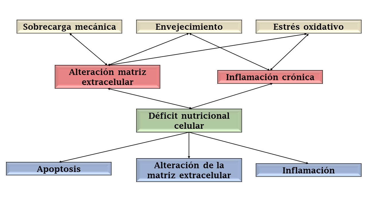
Así como ocurre en la artrosis, en todas las enfermedades crónicas y degenerativas ocurre la alteración del microambiente celular la cual en algunos causas es causa de la enfermedad, en otros es el origen de los síntomas y de las complicaciones que derivan de ella.
Contribuciones de la apiterapia a la regulación y recomposición del microambiente celular
La apiterapia es una herramienta terapéutica que produce modificaciones interesantes en el microambiente celular sano y enfermo (en la búsqueda del restablecimiento del ambiente natural).
Los mecanismos a través de los cuales la apiterapia contribuye a la regulación del microambiente celular son cada vez más conocidos:
Modulación del factor nuclear Kappa Beta. La activación del factor nuclear kappa beta ocurre en todos los procesos inflamatorios. Su activación crónica esta relacionada con múltiples enfermedades, desde las enfermedades autoinmunes hasta la enfermedad cardiovascular, es decir, la ausencia de su regulación conduce a alteraciones celulares propias de las enfermedades crónicas (29). Los productos de la colmena como el veneno de abejas, propóleo y jalea real modulan la activación del factor nuclear kappa Beta disminuyendo los efectos que se producen como consecuencia de la pérdida en su regulación (30–33).
Reducción del proceso inflamatorio. Los productos de la colmena inducen la disminución del proceso inflamatorio por regulación de la función de linfocitos y macrófagos, modulación de la actividad del sistema nervioso central y periférico y disminución de la liberación de las citoquinas proinflamatorias (34–36).
Incremento de la supervivencia celular. El veneno de abejas y jalea real incrementan la supervivencia celular mediante modulación de la vía de las caspasas, función mitocondrial y la prevención del daño del ADN (37–39).
Control del estrés oxidativo. La miel, polen y propóleos empleados en apiterapia poseen efecto secuestrante sobre los radicales libres mitigando así el daño celular y estructural inducido por el estrés oxidativo (40–44).
Inducción del metabolismo celular. La miel de abejas posee actividad regulatoria del metabolismo celular mediante la modulación de los receptores hormonales disponibles, particularmente del receptor para la insulina y el factor de crecimiento similar a la insulina (45).
Regulación del aporte de nutrientes en la célula. Tres efectos importantes de la miel y polen de abejas en este punto: regulación de la función mitocondrial, aporte directo de minerales, aminoácidos y vitaminas e incremento en la captación de los nutrientes disponibles en el microambiente celular (46,47).
Modulación de la liberación de factores de crecimiento. El veneno de abejas modula el efecto de los factores de crecimiento transformantes previniendo el reemplazo del tejido por fibra no funcional (48).
Disminución de la velocidad de envejecimiento celular. La jalea real previene la entrada en senescencia de distintos grupos celulares (49). El consumo regular de productos de la colmena como la miel, polen y propóleo reduce la velocidad con la cual se produce el acortamiento de los telómeros, indicador de envejecimiento celular (50).
Es por estos motivos que la apiterapia se convierte en una herramienta terapéutica útil y necesaria en el tratamiento de las condiciones que cursan con alteraciones del microambiente celular y, en todos los casos, potenciando el efecto regulador de otras terapéuticas.
Si te gustó este artículo te invitamos a compartirlo y suscribirte en el link «Newsletter» para recibir más información y contenidos relacionados
Referencias bibliográficas
1. Rehfeldt F, Engler AJ, Eckhardt A, Ahmed F, Discher DE. Cell responses to the mechanochemical microenvironment-Implications for regenerative medicine and drug delivery. Advanced Drug Delivery Reviews. 2007.
2. Moore KA, Lemischka IR. Stem cells and their niches. Science. 2006.
3. Freitas-Rodríguez S, Folgueras AR, López-Otín C. The role of matrix metalloproteinases in aging: Tissue remodeling and beyond. Biochim Biophys Acta – Mol Cell Res [Internet]. 2017 Nov;1864(11):2015–25. Available from: https://linkinghub.elsevier.com/retrieve/pii/S0167488917301180
4. Chanmee T, Ontong P, Itano N. Hyaluronan: A modulator of the tumor microenvironment. Cancer Lett [Internet]. 2016 May;375(1):20–30. Available from: https://linkinghub.elsevier.com/retrieve/pii/S0304383516300994
5. Zhuang X, Zhang H, Hu G. Cancer and Microenvironment Plasticity: Double-Edged Swords in Metastasis. Trends Pharmacol Sci [Internet]. 2019 Jun;40(6):419–29. Available from: https://linkinghub.elsevier.com/retrieve/pii/S016561471930080X
6. Greten FR, Grivennikov SI. Inflammation and Cancer: Triggers, Mechanisms, and Consequences. Immunity [Internet]. 2019 Jul;51(1):27–41. Available from: https://linkinghub.elsevier.com/retrieve/pii/S107476131930295X
7. Qu F, Guilak F, Mauck RL. Cell migration: implications for repair and regeneration in joint disease. Nat Rev Rheumatol [Internet]. 2019 Mar 7;15(3):167–79. Available from: http://www.nature.com/articles/s41584-018-0151-0
8. Wu T, Dai Y. Tumor microenvironment and therapeutic response. Cancer Lett [Internet]. 2017 Feb;387:61–8. Available from: https://linkinghub.elsevier.com/retrieve/pii/S0304383516300155
9. Hirata E, Sahai E. Tumor Microenvironment and Differential Responses to Therapy. Cold Spring Harb Perspect Med [Internet]. 2017 Jul;7(7):a026781. Available from: http://perspectivesinmedicine.cshlp.org/lookup/doi/10.1101/cshperspect.a026781
10. Bhat V, Allan AL, Raouf A. Role of the Microenvironment in Regulating Normal and Cancer Stem Cell Activity: Implications for Breast Cancer Progression and Therapy Response. Cancers (Basel) [Internet]. 2019 Aug 24;11(9):1240. Available from: https://www.mdpi.com/2072-6694/11/9/1240
11. Weinberg F, Ramnath N, Nagrath D. Reactive Oxygen Species in the Tumor Microenvironment: An Overview. Cancers (Basel) [Internet]. 2019 Aug 16;11(8):1191. Available from: https://www.mdpi.com/2072-6694/11/8/1191
12. Gobaa S, Gayet R V., Lutolf MP. Artificial niche microarrays for identifying extrinsic cell-fate determinants. In 2018. p. 51–69. Available from: https://linkinghub.elsevier.com/retrieve/pii/S0091679X1830075X
13. Mitchell JP, Carmody RJ. NF-κB and the Transcriptional Control of Inflammation. In 2018. p. 41–84. Available from: https://linkinghub.elsevier.com/retrieve/pii/S1937644817300825
14. Hoffman BD. The Detection and Role of Molecular Tension in Focal Adhesion Dynamics. In 2014. p. 3–24. Available from: https://linkinghub.elsevier.com/retrieve/pii/B9780123946249000014
15. Obinata M, Yanai N. Cellular and molecular regulation of an erythropoietic inductive microenvironment (EIM). Cell Struct Funct [Internet]. 1999 Aug;24(4):171–9. Available from: http://www.ncbi.nlm.nih.gov/pubmed/10532351
16. Goldring MB, Berenbaum F. The regulation of chondrocyte function by proinflammatory mediators: prostaglandins and nitric oxide. Clin Orthop Relat Res [Internet]. 2004 Oct;(427 Suppl):S37-46. Available from: http://www.ncbi.nlm.nih.gov/pubmed/15480072
17. Kong HJ, Mooney DJ. Microenvironmental regulation of biomacromolecular therapies. Nat Rev Drug Discov [Internet]. 2007 Jun;6(6):455–63. Available from: http://www.ncbi.nlm.nih.gov/pubmed/17541418
18. Portella L, Scala S. Ionizing radiation effects on the tumor microenvironment. Semin Oncol [Internet]. 2019 Jul; Available from: https://linkinghub.elsevier.com/retrieve/pii/S0093775419300971
19. Koelwyn GJ, Quail DF, Zhang X, White RM, Jones LW. Exercise-dependent regulation of the tumour microenvironment. Nat Rev Cancer [Internet]. 2017 Oct 1;17(10):620–32. Available from: http://www.nature.com/articles/nrc.2017.78
20. Luo G, Liu N. An integrative theory for cancer (Review). Int J Mol Med [Internet]. 2018 Nov 28; Available from: http://www.spandidos-publications.com/10.3892/ijmm.2018.4004
21. Wang T, Nasser MI, Shen J, Qu S, He Q, Zhao M. Functions of Exosomes in the Triangular Relationship between the Tumor, Inflammation, and Immunity in the Tumor Microenvironment. J Immunol Res [Internet]. 2019 Aug 1;2019:1–10. Available from: https://www.hindawi.com/journals/jir/2019/4197829/
22. Wawman RE, Bartlett H, Oo YH. Regulatory T Cell Metabolism in the Hepatic Microenvironment. Front Immunol [Internet]. 2018 Jan 8;8. Available from: http://journal.frontiersin.org/article/10.3389/fimmu.2017.01889/full
23. Yurdagul A, Orr AW. Blood Brothers: Hemodynamics and Cell–Matrix Interactions in Endothelial Function. Antioxid Redox Signal [Internet]. 2016 Sep;25(7):415–34. Available from: http://www.liebertpub.com/doi/10.1089/ars.2015.6525
24. Song R, Hu X-Q, Zhang L. Glucocorticoids and programming of the microenvironment in heart. J Endocrinol [Internet]. 2019 Jul;242(1):T121–33. Available from: https://joe.bioscientifica.com/view/journals/joe/242/1/JOE-18-0672.xml
25. McGarry T, Biniecka M, Veale DJ, Fearon U. Hypoxia, oxidative stress and inflammation. Free Radic Biol Med [Internet]. 2018;125:15–24. Available from: http://www.ncbi.nlm.nih.gov/pubmed/29601945
26. Morgillo F, Dallio M, Della Corte CM, Gravina AG, Viscardi G, Loguercio C, et al. Carcinogenesis as a Result of Multiple Inflammatory and Oxidative Hits: a Comprehensive Review from Tumor Microenvironment to Gut Microbiota. Neoplasia [Internet]. 2018 Jul;20(7):721–33. Available from: https://linkinghub.elsevier.com/retrieve/pii/S1476558618301246
27. Zhang Y, Liu Y, Liu H, Tang WH. Exosomes: biogenesis, biologic function and clinical potential. Cell Biosci [Internet]. 2019 Dec 15;9(1):19. Available from: https://cellandbioscience.biomedcentral.com/articles/10.1186/s13578-019-0282-2
28. Williams R, Kalita D. A Physician’s Handbook on Orthomolecular Medicine. Primera ed. New York: Pergamon press; 1977.
29. Tornatore L, Thotakura AK, Bennett J, Moretti M, Franzoso G. The nuclear factor kappa B signaling pathway: integrating metabolism with inflammation. Trends Cell Biol [Internet]. 2012 Nov;22(11):557–66. Available from: http://linkinghub.elsevier.com/retrieve/pii/S0962892412001407
30. Shin S, Ye M, Choi S, Park K. Anti-inflammatory effect of bee venom in an allergic chronic rhinosinusitis mouse model. Mol Med Rep [Internet]. 2018 Mar 9; Available from: http://www.spandidos-publications.com/10.3892/mmr.2018.8720
31. Choi K, Hwang C, Gu S, Park M, Kim J, Park J, et al. Cancer Cell Growth Inhibitory Effect of Bee Venom via Increase of Death Receptor 3 Expression and Inactivation of NF-kappa B in NSCLC Cells. Toxins (Basel) [Internet]. 2014 Jul 25;6(8):2210–28. Available from: http://www.mdpi.com/2072-6651/6/8/2210
32. Yang X-Y, Yang D, Wei-Zhang, Wang J-M, Li C-Y, Hui-Ye, et al. 10-Hydroxy-2-decenoic acid from Royal jelly: a potential medicine for RA. J Ethnopharmacol [Internet]. 2010 Mar 24;128(2):314–21. Available from: http://www.ncbi.nlm.nih.gov/pubmed/20138211
33. Nie J, Chang Y, Li Y, Zhou Y, Qin J, Sun Z, et al. Caffeic Acid Phenethyl Ester (Propolis Extract) Ameliorates Insulin Resistance by Inhibiting JNK and NF-κB Inflammatory Pathways in Diabetic Mice and HepG2 Cell Models. J Agric Food Chem [Internet]. 2017 Oct 18;65(41):9041–53. Available from: https://pubs.acs.org/doi/10.1021/acs.jafc.7b02880
34. KIM J-Y, LEE W-R, KIM K-H, AN H-J, CHANG Y-C, HAN S-M, et al. Effects of bee venom against Propionibacterium acnes-induced inflammation in human keratinocytes and monocytes. Int J Mol Med [Internet]. 2015 Jun;35(6):1651–6. Available from: https://www.spandidos-publications.com/10.3892/ijmm.2015.2180
35. Ali M. Studies on Bee Venom and Its Medical Uses. Int J Adv Res …. 2012;1:1–15.
36. Pasupuleti VR, Sammugam L, Ramesh N, Gan SH. Honey, Propolis, and Royal Jelly: A Comprehensive Review of Their Biological Actions and Health Benefits. Oxid Med Cell Longev [Internet]. 2017;2017:1259510. Available from: http://www.ncbi.nlm.nih.gov/pubmed/28814983
37. Takahashi Y, Hijikata K, Seike K, Nakano S, Banjo M, Sato Y, et al. Effects of Royal Jelly Administration on Endurance Training-Induced Mitochondrial Adaptations in Skeletal Muscle. Nutrients [Internet]. 2018 Nov 12;10(11):1735. Available from: http://www.mdpi.com/2072-6643/10/11/1735
38. Jung S, Lee K-W, Choi S-M, Yang E. Bee Venom Protects against Rotenone-Induced Cell Death in NSC34 Motor Neuron Cells. Toxins (Basel) [Internet]. 2015 Sep 21;7(9):3715–26. Available from: http://www.mdpi.com/2072-6651/7/9/3715
39. Almeer RS, AlBasher GI, Alarifi S, Alkahtani S, Ali D, Abdel Moneim AE. Royal jelly attenuates cadmium-induced nephrotoxicity in male mice. Sci Rep [Internet]. 2019 Dec 9;9(1):5825. Available from: http://www.nature.com/articles/s41598-019-42368-7
40. Khalil MI, Tanvir EM, Afroz R, Sulaiman SA, Gan SH. Cardioprotective Effects of Tualang Honey: Amelioration of Cholesterol and Cardiac Enzymes Levels. Biomed Res Int [Internet]. 2015;2015:1–8. Available from: http://www.hindawi.com/journals/bmri/2015/286051/
41. Prior RL, Cao G. In vivo total antioxidant capacity: comparison of different analytical methods. Free Radic Biol Med [Internet]. 1999 Dec;27(11–12):1173–81. Available from: http://www.ncbi.nlm.nih.gov/pubmed/10641708
42. Bankova V, Galabov AS, Antonova D, Vilhelmova N, Di Perri B. Chemical composition of Propolis Extract ACF® and activity against herpes simplex virus. Phytomedicine [Internet]. 2014 Sep;21(11):1432–8. Available from: http://linkinghub.elsevier.com/retrieve/pii/S0944711314002104
43. Fratellone PM, Tsimis F, Fratellone G. Apitherapy Products for Medicinal Use. J Altern Complement Med. 2016;5(6):6–7.
44. Campos MGR, Bogdanov S, de Almeida-Muradian LB, Szczesna T, Mancebo Y, Frigerio C, et al. Pollen composition and standardisation of analytical methods. Vol. 47, Journal of Apicultural Research. 2008. p. 154–61.
45. Lori G, Cecchi L, Mulinacci N, Melani F, Caselli A, Cirri P, et al. Honey extracts inhibit PTP1B, upregulate insulin receptor expression, and enhance glucose uptake in human HepG2 cells. Biomed Pharmacother [Internet]. 2019 May;113:108752. Available from: https://linkinghub.elsevier.com/retrieve/pii/S0753332218346948
46. Ricigliano VA, Fitz W, Copeland DC, Mott BM, Maes P, Floyd AS, et al. The impact of pollen consumption on honey bee ( Apis mellifera ) digestive physiology and carbohydrate metabolism. Arch Insect Biochem Physiol [Internet]. 2017 Oct;96(2):e21406. Available from: http://doi.wiley.com/10.1002/arch.21406
47. Afrin S, Gasparrini M, Forbes-Hernández TY, Cianciosi D, Reboredo-Rodriguez P, Manna PP, et al. Protective effects of Manuka honey on LPS-treated RAW 264.7 macrophages. Part 1: Enhancement of cellular viability, regulation of cellular apoptosis and improvement of mitochondrial functionality. Food Chem Toxicol [Internet]. 2018 Nov;121:203–13. Available from: https://linkinghub.elsevier.com/retrieve/pii/S0278691518306379
48. Lee W-R, Pak S, Park K-K. The Protective Effect of Bee Venom on Fibrosis Causing Inflammatory Diseases. Toxins (Basel) [Internet]. 2015 Nov 16;7(12):4758–72. Available from: http://www.mdpi.com/2072-6651/7/11/4758
49. Jiang C, Liu X, Li C, Qian H, Chen D, Lai C, et al. Anti-senescence effect and molecular mechanism of the major royal jelly proteins on human embryonic lung fibroblast (HFL-I) cell line. J Zhejiang Univ B [Internet]. 2018 Dec 17;19(12):960–72. Available from: http://link.springer.com/10.1631/jzus.B1800257
50. Nasir NFM, Kannan TP, Sulaiman SA, Shamsuddin S, Azlina A, Stangaciu S. The relationship between telomere length and beekeeping among Malaysians. Age (Omaha). 2015;37(3).